Multimodal Neurophysiology
Project leader: Prof. Dr. Michèle Hubli
The spinothalamic system transmits noxious and innocuous thermal and noxious mechanical information. While the assessment of thermo-nociceptive pathways using contact heat evoked potentials (CHEPs) is well established in diagnosis of incomplete spinal cord lesions, objective readouts for pinprick evoked pain (pinprick evoked potentials / PEPs) and cold evoked potentials (CEPs) are more novel.
Neurophysiological techniques are crucial in the early diagnosis of nerve conduction impairments. Here we want to investigate if multimodal neurophysiology improves the clinical diagnosis of myelopathy often presented high variability and subtle clinical presentations. In addition, we aim to assess the integrity of the spinothalamic tract by examining sub-modality specificity of various evoked brain potentials and quantitative sensory testing.
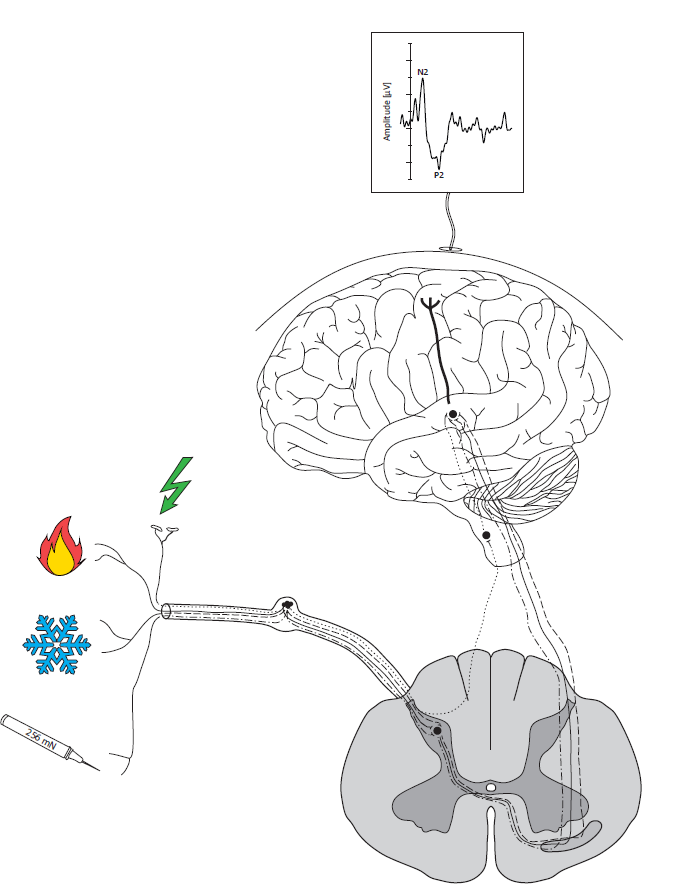
Figure 1: Multimodal neurophysiology to assess the integrity of the spinothalamic tract and the dorsal column. Our multimodal neurophysiological test battery comprises somatosensory evoked potentials (non-noxious electrical stimuli), contact heat evoked potentials (heat stimuli), cold evoked potentials (innocuous cold stimuli) and pinprick evoked potentials (mechanical stimuli).
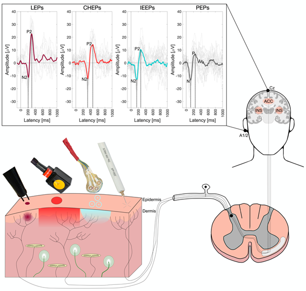
Figure 2: Pain-related evoked potentials (PREPs). PREPs can be recorded from the vertex (Cz) after different types of stimulation activating small-diameter fibers in the skin. Radiant (laser) or contact heat activate thermo-nociceptive fibers, pinprick stimulation activated mechano-nociceptive fibers, and intra-epidermal electrical stimulation activates free nerve endings in the epidermis. Once the afferent volley is conveyed into the dorsal horn it ascends within the contralateral spinothalamic tract to the brain. Typical cortical components of PREPs recorded from the vertex are shown in the top panel: Aδ-fiber mediated thermally evoked potential (LEPs and CHEPs) consists of large N2-P2 complexes. CHEPs have longer latencies and smaller amplitudes than LEPs, but CHEPs amplitudes can be increased with appropriate stimulation protocols ((Kramer, Haefeli, Jutzeler, Steeves, & Curt, 2013). Intra-epidermal electrical stimulation evoked potentials (IEEPs) using a triple concentric planar electrode (Lutolf, Julio, Schubert, & Hubli, 2022) show similar waveforms to LEPs and CHEPs, but a shorter latency and often smaller amplitudes. Importantly, IES induces less painful sensation than laser or contact heat stimulation. Pinprick evoked potentials (PEPs) show the fastest latencies and lowest amplitudes due to the least synchronous afferent volley (often induced by manual pinprick application) and potential co-activation of mechanosensitive Aβ-fibers (Rosner, Scheuren, Stalder, Curt, & Hubli, 2020) (created by Dr. S Uldry).
Project partners:
Prof. Dr. Kip Kramer, International Collaboration On Repair Discoveries (ICORD), University of British Columbia, Vancouver, British Columbia, Canada
Prof. Dr. med. Jan Rosner, Danish Pain Research Center, Department of Clinical Medicine, Aarhus University, Aarhus, Denmark
PD Dr. med. Philipp Hüllemann, Division of Neurological Pain Research and Therapy, Department of Neurology, University Hospital Schleswig-Holstein, Kiel, Germany
PD Dr. med. Martin Schubert, Spinal Cord Injury Center, Balgrist University Hospital, University of Zurich, Switzerland
Publications:
- Uldry S, Schneuwly M, Scheuren PS, Pfender N, Zipser C, Hubli M, Schubert M. Intra-epidermal electrically evoked potentials are sensitive to detect degenerative cervical myelopathy suggesting their spinothalamic propagation. Clin Neurophysiol. 2024 (accepted).
- Scheuren P, Nauer N, Rosner R, Curt A, Hubli M. Cold evoked potentials elicited by rapid cooling of the skin across different age groups and stimulation sites. Sci Rep. 2022 Mar 9;12(1):4137.
- Lütolf R, Júlio SU, Schubert M, Hubli M. Intra-epidermal evoked potentials: A promising tool for spinal disorders? Neurophysiol Clin. 2021 Dec 22: S0987-7053(21)00125-8.
- Scheuren PS, David G, Kipling Kramer JL, Jutzeler CR, Hupp M, Freund P, Curt A, Hubli M*, Rosner J. Combined Neurophysiologic and Neuroimaging Approach to Reveal the Structure-Function Paradox in Cervical Myelopathy. Neurology. 2021 Oct 11;97(15):e1512-e1522. (*shared last authorship).
- Jutzeler CR, Linde LD, Rosner J, Hubli M, Curt A, Kramer JLK. Single-trial averaging improves the physiological interpretation of contact heat evoked potentials. Neuroimage. 2021 Jan 15; 225:117473.
- De Schoenmacker I, Berry C, Blouin JS, Rosner J, Hubli M, Jutzeler CR, Kramer JLK. An intensity matched comparison of laser- and contact heat evoked potentials. Sci Rep. 2021 Mar 25;11(1):6861.
- Rosner J, Scheuren PS, Stalder S, Curt A, Hubli M. Pinprick-evoked potentials: Moving towards a clinical assessment tool. Pain Med. 2020 Apr 1;21(4):736-746.
- Rosner J, Rinert J, Ernst M, Curt A, Hubli M. Cold evoked potentials: Acquisition from cervical dermatomes. Neurophys Clin. 2019 Feb;49(1):49-57.
- Rosner J, Sirucek L, Hostettler P, Scheuren PS, Rinert J, Curt A, Jutzeler CR, Hubli M. Normative data of contact heat evoked potentials from lower extremities. Sci Rep. 2018 Jul;8(1):11003.
- Rosner J*, Hubli M*, Hostettler P, Scheuren PS, Rinert J, Kramer JLK, Hupp M, Curt A, Jutzeler CR. Contact heat evoked potentials: reliable acquisition from lower extremities. Clin Neurophysiol. 2018;129(3): 584-591. (*shared first authors).
For further information please contact: Prof. Dr. Michèle Hubli